No fluff. No fads. Deep-dive investigative reports from the surgeon who actually sees the inside of the joints.
If your kneecap has shifted, slipped, or fully popped out — even once — you are not "just flexible." You have an anatomy problem that rarely fixes itself, and the longer you wait, the more cartilage it costs you.
The patella is a floating bone. It rides in a shallow groove on the front of the femur called the trochlear groove, held in place by a tiny ligament on the inside of the knee — the medial patellofemoral ligament, or MPFL. When the knee bends and straightens, the kneecap slides up and down that groove like a train on a track.
Dislocation happens when the patella jumps the track. Almost always it shifts laterally (toward the outside of the knee). In the process, it tears the MPFL and often shears a piece of cartilage off the back of the kneecap or the edge of the femur. That cartilage piece — called an osteochondral fragment — is often the real long-term problem.
Most patients remember the pop. Few realize that a single dislocation can leave a dime-sized divot of cartilage floating in the joint. That is the injury that ages a knee by twenty years.
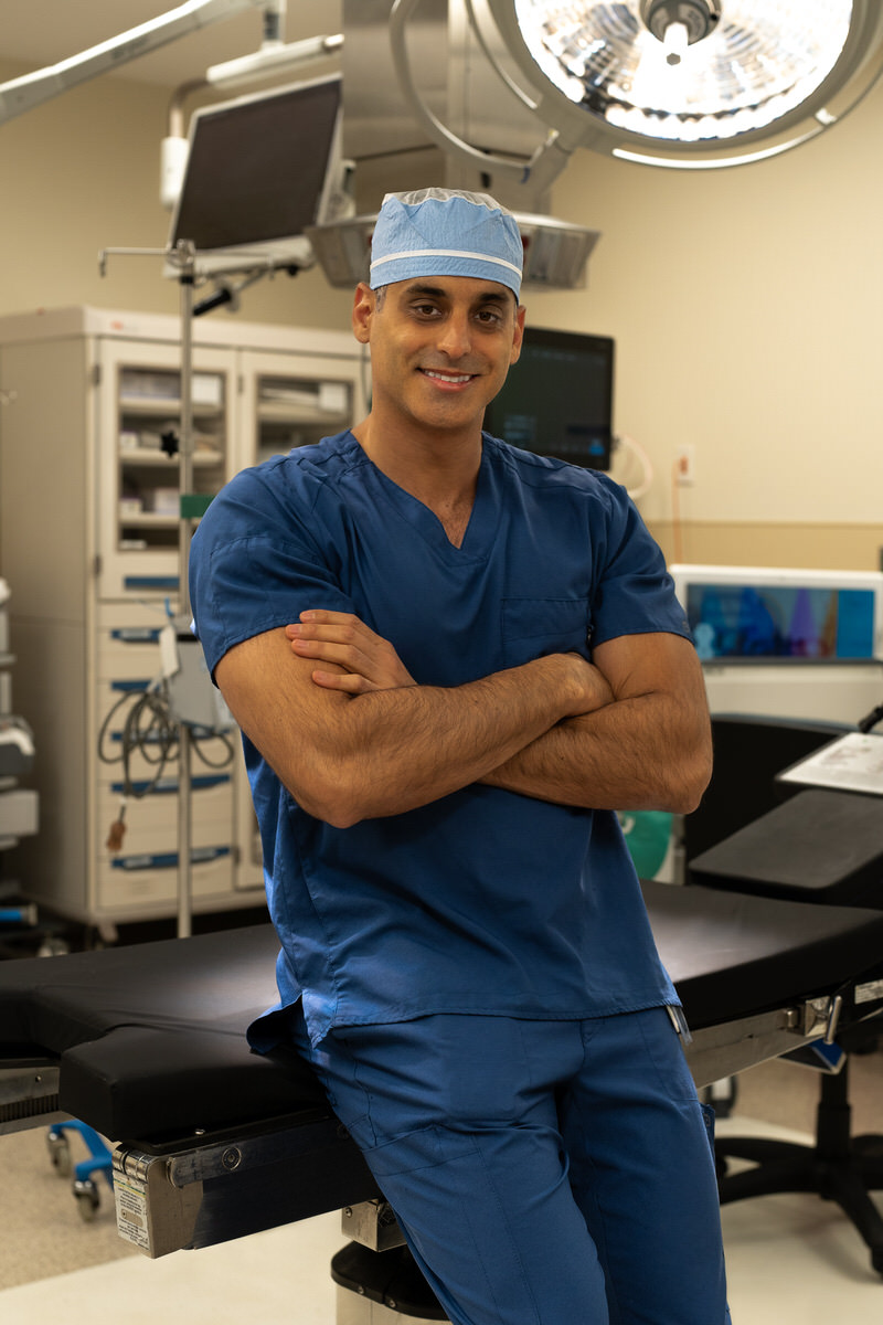
Dr. Sameh Elguizaoui, M.D. — Board-Certified Orthopedic Surgeon, Sports Medicine SpecialistA first-time dislocation is not automatically a surgical problem. The priority in the first six weeks is precise: rule out loose cartilage, protect the torn MPFL, and rebuild quad strength before the kneecap gets lazy.
X-rays miss most cartilage fragments. A high-quality MRI is mandatory after a first dislocation to find loose pieces and grade the MPFL tear.
A patellar-stabilizing brace during ambulation for 4 weeks. Full passive range of motion early — frozen knees do not do well.
The vastus medialis (the teardrop on the inside of the quad) is the patella's strongest dynamic stabilizer. Specific PT targets VMO recovery first.
Recurrence is not bad luck. The anatomy that let the kneecap dislocate the first time is still there. The MPFL has healed loose, the groove is still shallow, the quad is still weaker than it was before the injury. Each subsequent dislocation is easier to produce, and each one chips off more cartilage.
By the second dislocation, the math changes. The question is no longer can we rehab this — it is which reconstruction stops the cycle.
Medial patellofemoral ligament reconstruction is the workhorse procedure for recurrent patellar instability. It is an outpatient arthroscopic-assisted surgery that rebuilds the checkrein that keeps the kneecap from sliding laterally.
A strip of the gracilis tendon (a low-demand hamstring) is harvested through a half-inch incision, or a donor allograft is used.
Using fluoroscopic landmarks, a small tunnel is drilled at the native MPFL footprint on the femur. Two small sockets go into the medial edge of the patella.
The graft is passed and tensioned with the knee at 30° flexion — the position the native MPFL is tightest. Tension is a matter of millimeters; this is where surgeon experience shows.
If TT-TG distance is > 20 mm, we add a tibial tubercle osteotomy in the same sitting. If there is a cartilage divot, we fix it now — not later.
Soft tissue
Bone realignment
A brace prevents some dislocations. It does not rebuild a torn ligament, it does not protect the cartilage you lose with each new event, and it does not let you return to cutting sports with confidence. For recurrent dislocators, bracing is a bridge — not a destination.
Patellar instability is often bilateral in its anatomy even when only one side has dislocated. If you have high-risk features on MRI, preventive PT on the asymptomatic side is worth the effort.
MPFL reconstruction can be performed with physeal-sparing techniques in adolescents with open growth plates. The anatomy of the pediatric knee changes the graft path but not the principle. Timing is individualized to skeletal maturity.
Left knee: typically within a week once you are off opioids. Right knee: 4–6 weeks, when you have the quad strength to slam a brake pedal without hesitation.
We address both at the same surgery. Cartilage that is ignored at the index procedure is the single biggest predictor of a bad long-term outcome. See our cartilage restoration deep dive for the biologics options.
Get Started
Take the first step toward recovery. Schedule a consultation with Dr. Elguizaoui to discuss your condition and explore your treatment options.